Invented by Roger A. Sabbadini, William A. Garland, Genevieve Hansen, Steven Tarran Jones, David Gareth Williams, Apollo Endosurgery Inc
The development of humanized antibodies for targeting S1P has opened up new possibilities for treating these diseases. By specifically binding to S1P, these antibodies can modulate immune cell migration and function, leading to potential therapeutic benefits. Several humanized antibodies targeting S1P have already been approved by regulatory authorities and are being used in clinical practice.
One of the key drivers of the market for humanized antibodies targeting S1P is the increasing prevalence of diseases where S1P dysregulation is implicated. For example, multiple sclerosis affects millions of people worldwide, and the development of effective therapies is a major unmet need. Humanized antibodies targeting S1P have shown promising results in clinical trials for multiple sclerosis, providing hope for patients and driving the demand for these therapies.
Additionally, the market is also driven by the growing understanding of the role of S1P in other diseases, such as rheumatoid arthritis and cancer. Rheumatoid arthritis is a chronic autoimmune disease that affects the joints, and S1P has been found to play a role in the migration and activation of immune cells involved in the disease process. Humanized antibodies targeting S1P have shown potential in reducing disease activity and improving symptoms in patients with rheumatoid arthritis.
In the field of oncology, S1P has been implicated in tumor growth, angiogenesis, and metastasis. Humanized antibodies targeting S1P have shown promise in preclinical and early clinical studies for various types of cancer, including breast cancer and melanoma. The potential of these therapies to improve patient outcomes in cancer treatment has generated significant interest from both pharmaceutical companies and investors.
The market for humanized antibodies targeting S1P is also driven by advancements in antibody engineering and manufacturing technologies. The development of novel antibody formats, such as bispecific antibodies and antibody-drug conjugates, allows for improved targeting and delivery of therapeutic payloads to the desired sites of action. Furthermore, advancements in manufacturing technologies, such as cell line development and purification processes, have increased the scalability and cost-effectiveness of producing these antibodies, making them more accessible to patients.
However, the market for humanized antibodies targeting S1P also faces challenges. The high cost of developing and manufacturing these therapies, coupled with the stringent regulatory requirements for approval, can limit their accessibility to patients. Additionally, the competitive landscape is becoming increasingly crowded, with several companies developing their own humanized antibodies targeting S1P. This competition can make it challenging for new entrants to gain market share.
In conclusion, the market for humanized antibodies and compositions for binding S1P is experiencing significant growth due to the increasing prevalence of diseases where S1P dysregulation is implicated. The potential of these therapies to provide targeted and effective treatments for diseases such as multiple sclerosis, rheumatoid arthritis, and cancer has generated significant interest from both the pharmaceutical industry and investors. However, challenges such as high development costs and competition in the market need to be addressed to ensure the continued growth and accessibility of these therapies.

The Apollo Endosurgery Inc invention works as follows
The present invention is a new anti-S1P agent, namely humanized monoclonal antibody (and antigen binding fractions thereof), which are specifically reactive with S1P. It also relates to compositions that contain such antibodies or fragments, and to the use of these antibodies (or their fragments) to treat conditions and diseases associated with abnormal levels of S1P.
Background for Humanized antibodies and compositions for binding sphingosine-1-phosphate
1. “1.
The present invention relates to agents that bind sphingosine-1-phosphate (S1P), particularly to humanized monoclonal antibodies, antibody fragments, and antibody derivatives specifically reactive to S1P under physiological conditions. These agents can be used to treat and/or prevent various diseases or disorders by delivering pharmaceutical compositions containing such agents.
The following description contains information that could be helpful in understanding the invention. This is not to say that the information or publications referred to herein are prior art or particularly relevant for the invention claimed.
2. Background
Bioactive Signaling Lipids
Lipids, and their derivatives, are now recognized as an important target for medical research. They are not just structural elements of cell membranes, or a source for energy in?-oxidation or glycolysis, or for other metabolic processes. Certain bioactive lipids are important signaling mediators in both animal and human diseases. Most lipids in the plasma membrane are structural, but a small percentage of them is involved in relaying external stimuli to cells. ?Lipid signaling? Lipid signaling can refer to a variety of cell signal transduction pathways which use lipids in the membrane of cells as second messengers. It may also refer to a direct interaction between a lipid-signaling molecule and its receptor. Lipid signaling pathways can be activated by extracellular stimuli ranging from cytokines to growth factors. They regulate cell fate decisions, such as apoptosis and differentiation. As more bioactive molecules are discovered and their effects characterized, the research into bioactive signaling is a field of intense scientific study.
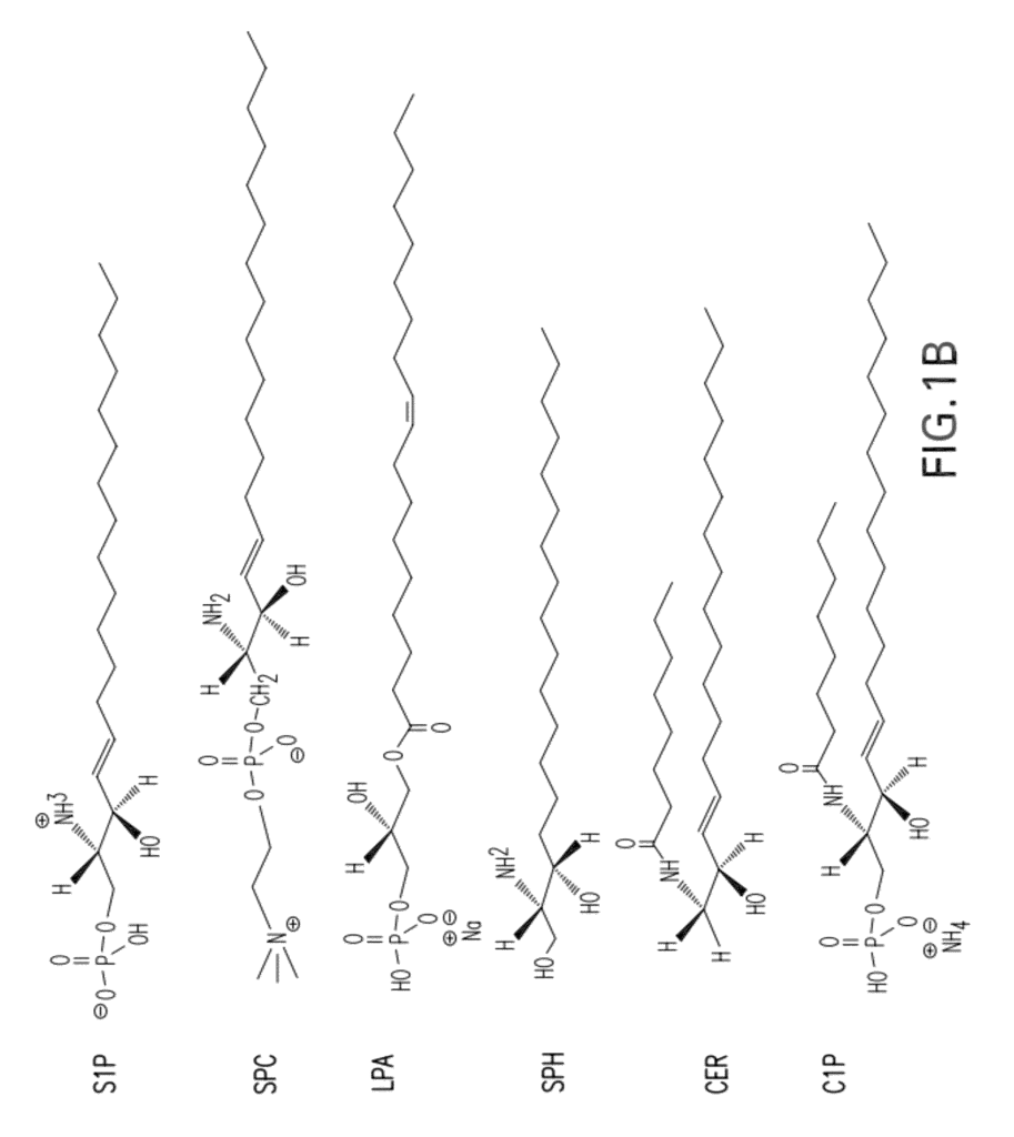
Bioactive lipids are eicosanoids such as cannabinoids and leukotrienes. They also include phospholipids (such as phosphatidic and phosphatidylglycerol), cardiolipins (such as lysophosphatidylcholine and LPA), and lysophospholipids (such as lysophosphatidylcholine and LPA). Bioactive signaling lipid mediators also include the sphingolipids such as sphingomyelin, ceramide, ceramide-1-phosphate, sphingosine, sphingosylphosphoryl choline, sphinganine, sphinganine-1-phosphate (Dihydro-S1P) and sphingosine-1-phosphate. Sphingolipids, and their derivatives, are a grouping of intracellular and extracellular signaling molecules that have pleiotropic impacts on important cellular functions. Other examples of bioactive signaling lipids include phosphatidylserine (PS), phosphatidylinositol (PI), phosphatidylethanolamine (PEA), diacylglyceride (DG), sulfatides, gangliosides, and cerebrosides.
Sphingolipids were originally named after the Sphinx due to their initial mystery. Sphingolipids are a unique class of lipids that were named after the Sphinx due to their initial mysterious nature. Recent studies have shown, however, that they also function as signaling and regulation molecules in cells (Hannun et.al., Advances in Lipid Research). Lipid Res. 25:27-41, 1993; Speigel, et al., FASEB J. 10:1388-1397, 1996; Igarashi, J. Biochem 122:1080-1087, 1997; Hla, T. (2004). Semin Cell Dev Biol 15, 513-2. Gardell, S. E. Dubin, A. E. and Chun, J. (2006). Trends Mol Med. 12, 65-75. Sphingolipids, which are the primary structural component of cell membranes and also act as cellular regulatory and signaling molecules (Hannun & Bell, Adv. Lipid Res. 25: 27-41, 1993; Igarashi, J. Biochem 122: 1080-1087, 1997). The sphingolipid signaling mediators, ceramide (CER), sphingosine (SPH) and sphingosine-1-phosphate (S1P), have been most widely studied and have recently been appreciated for their roles in the cardiovascular system, angiogenesis and tumor biology (Claus, et al., Curr Drug Targets 1: 185-205, 2000; Levade, et al., Circ. Res. 89: 957-968, 2001; Wang, et al., J. Biol. Chem. Wascholowski, Giannis and Drug News Perspect. 14: 581-90, 2001; Spiegel, S. & Milstien, S. (2003). Sphingosine-1-phosphate: an enigmatic signaling lipid. Nat Rev Mol Cell Biol 4, 397-407).
For a review on sphingolipid metabolic pathways, please see Liu et. al., Critical Rev. Clin. Lab. Sci. 36:511-573, 1999. Hannun et. al., Advances in Molecular Biology, vol. Lipid Res. 25:27-41, 1993; Liu, et al., Crit. Rev. Clin. Lab. Sci. 36:511-573, 1999; Igarashi, J. Biochem. 122:1080-1087, 1997; Oral, et al., J. Biol. Chem. Chem.
S1P mediates cell proliferation and protects against apoptosis by activating survival pathways” (Maceyka et al. (2002), BBA, vol. 1585): 192-201, and Spiegel, et al. Nature Reviews Molecular Cell Biology vol. 4: 397-407). The balance between CER/SPH and S1P is thought to be a rheostat that determines whether or not a cell will enter the death pathway, or if it will be protected from apoptosis. Sphingosine Kinase (SPHK), the key regulator of the rheostat, converts the death-promoting signaling lipids CER/SPH into growth-promoting S1P. The fate of S1P is twofold: it can be degraded either by S1P Lyase (an enzyme that breaks S1P down into phosphoethanolamine, hexadecanal or less commonly, by S1P Phosphatase) to SPH.
![]()
The pleiotropic activities of S1P is mediated by a family G protein-coupled (GPCR) receptors originally known as Endothelial Differentiation Genes” (EDG). Five GPCRs were identified as S1P high-affinity receptors (S1PRs), namely S1P1/EDG-1 (formerly known as Endothelial Differentiation Genes (EDG)), S1P2/EDG-5 (formerly known as Endothelial Differentiation Genes (EDG)), S1P3/EDG-3 (formerly known as S1P3/EDG-3), S1P4/EDG-6 (formerly S1P5/EDG-8), S1P, EDG), S1P), S1P), S1P), S1P), S1P), S1P) and S1P5/EDG), S1P, S1P) S1P), S1P5/EDG) (Lee EDG), S1P (S1P) (Lee EDG8), S1P8 (Lee EDG), S1P), S1P1 (S1P1/EDG), S1P2/EDG-5 (EDG), S1P2/EDG-5 (EDG-5 S1P2/EDG-5 S1P3/EDG-3 S1P3 S1P3 S1P3 S1P3 S1P3 S1P3 S1P3 S1P3 S2 EDG5 S1P3 S1P3/EDG-3 S1P3 S1P3 S S1P elicits many responses that are coupled with heterotrimeric G proteins such as Gq-, G12-13, and Rho small GTPases (Gardell et al. 2006).
In the adult, S1P released from mast cells and platelets (Murata, et. al., 2000) creates a local pulse (sufficient to exceed the Kd for the S1PRs), which is used to promote wound healing and participate in the inflammatory reaction. In normal conditions, S1P levels in plasma are high (300-500nM). However, it is hypothesized that the majority of S1P could be “buffered”. The serum proteins (particularly lipoproteins, e.g. HDL>LDL>VLDL), and albumin may buffer the S1P, preventing it from activating S1PRs. This would lead to inappropriate inflammation and angiogenesis. There have been reports of S1P’s intracellular effects (see Spiegel S, Kolesnick (2002) in Leukemia vol. 16: 1596-602; Suomalainen, et al (2005), Am J Pathol, vol. 166: 773-81).
Widespread expression on the S1P receptors of cell surfaces allows S1P influence a spectrum of diverse cellular responses including proliferation, adhesion contraction, motility morphogenesis differentiation and survival. This response spectrum appears to be dependent on the expression patterns of S1P receptors in the cells and tissues. Recent studies have also shown that S1P can interact with growth factors, such as platelet-derived fibroblastic factor (bFGF), vascular enothelial graft growth factor(VEGF), or basic fibroblastic GF (bFGF). (2004), FASEB J, vol. 18: 341-3). S1P regulates a variety of cellular processes, including neuronal signals, wound healing and immune cell trafficking. It also affects reproduction, cardiovascular function, and reproduction. The alteration of S1P levels in these systems can cause a variety of pathophysiological conditions including cancer, inflammation and angiogenesis.
The treatment of many diseases and disorders including cardiovascular disease, cerebrovascular disease, and cancers involves reducing the levels of biologically-available S1P. This can be done alone or with other treatments. Although sphingolipid treatment strategies targeting key enzymes in the sphingolipid metabolism pathway, like SPHK, have already been proposed, the interference with S1P, the lipid mediator, has been neglected until recently, due to the difficulties of directly mitigating the S1P target. This is due, in part, to the difficulties of first raising and then detecting antibodies.
Recently, the production of antibodies specific to S1P was described. See, for example, the U.S. Patent Application Serial No. 20070148168; WO2007/053447. These antibodies can be used as molecular sponges for neutralizing extracellular S1P. They can, for instance, selectively adsorb S1P in serum. Also, see commonly owned U.S. Pat. Nos. Nos. 6,881,546 et 6,858,383 as well as U.S. Patent application Ser. No. 10/029,372. SPHINGOMAB, the murine monoclonal antigen (mAb), developed by Lpath, Inc., and described in some patents or applications listed above, was shown to be effective on models of human diseases. A humanized antibody is sometimes preferred to a murine one, especially for therapeutic purposes in humans where a human-antimouse antibodies (HAMAs) reaction may occur. This response can reduce the effectiveness by neutralizing binding activity or by clearing the antibody quickly from circulation. “The HAMA response may also lead to toxicities when mouse antibodies are administered in the future.
This article describes a humanized anti-S1P antigen. This antibody should have all of the benefits of a murine mAb, in terms of binding S1P to neutralize S1P to modulate disease states associated with S1P but none of the disadvantages when used in a hive context. This humanized antibody, also known as LT1009 (or sonepcizumab), has shown greater activity than the murine parent antibody in animal models.
3. Definitions
Before we describe the instant invention, we will define several terms that are used in the context. Other terms are also defined in the specification as needed. Terms of art will be used in this specification unless they are defined explicitly. If there is a conflict, this specification will prevail, including its definitions.
An ?immune-derived moiety? “An immune-derived moiety” includes any antibody (Ab), immunoglobulins (Ig), as well as any peptides, polypeptides, or fragments of these peptides or polypeptides that are capable of binding to an antigen or an epitope. Garland Publishing, 2001). The antigen in the present invention is a bioactive, lipid-based molecule.
An ?anti-S1P antibody? “Anti-S1P antibody” or “immune-derived moiety reacting against S1P” refers to any antibody or antibody-derived molecule that binds S1P. Any antibody or antibody-derived molecular that binds to S1P. These definitions will help you understand that antibodies or immune-derived molecules can be monoclonal, polyclonal, or generated in a variety ways. They may also be isolated from animals, including humans.
![]()
A ?bioactive lipid? Refers to a signaling lipid molecule. A bioactive lipid is not a part of the biological membrane that it signals from. This means, while a particular lipid species can exist in a certain place in a membrane (for instance, in a cell membrane or a membrane in an organelle), its signaling effect does not occur there. When a bioactive lipid is associated with a membrane, it’s not called a “bioactive lipid”. When associated with a biological membrane, it is not a “bioactive lipid” molecule. Bioactive lipids differ from structural lipids, such as membrane-bound phospholipids, in that they are involved in extracellular or intracellular signaling. They are also responsible for controlling the function of various types of cells, including differentiation, migration and proliferation, secretion and survival. Bioactive lipids are found in extracellular fluids in vivo. They can either be complexed (e.g., with serum proteins like albumin or lipoproteins) or in their ‘free’ form. Bioactive lipids can be found in extracellular fluids, where they are either complexed (e.g. with albumin and lipoproteins) or ‘free’, that is, uncomplexed. Some bioactive lipids act as extracellular mediators by activating membrane bound ion receptors or G-protein-coupled receptors. These, in turn activate complex signaling system that results in changes to cell function or survival. Bioactive lipids are intracellular mediators that can act by directly interacting intracellular components like enzymes and channels. LPA and S1P are two examples of bioactive fats.
Click here to view the patent on Google Patents.
Leave a Reply