Invented by Edward S. Boyden, Roderick A. Hyde, Muriel Y. Ishikawa, Edward K. Y. Jung, Nathan P. Myhrvold, Clarence T. Tegreene, Thomas A. Weaver, Charles Whitmer, Lowell L. Wood, JR., Victoria Y. H. Wood, Gearbox LLC
The Gearbox LLC invention works as follows
A method, a composition and a system that responds to ionizing radio waves to adjust biological activity.” In some cases, the ionizing energy is X-rays or extreme ultraviolet light that produce luminescent reactions that induce biologically reactive responses.

Background for Ionizing-radiation-responsive compositions, methods, and systems
In the detailed description that follows, the drawings are referred to. They form part of this document. In the drawings, symbols that are similar to one another usually identify similar components unless context dictates something else. The detailed description, drawings and claims do not limit the illustrative examples. Other embodiments can be used, and changes made without departing the spirit or scope presented here.
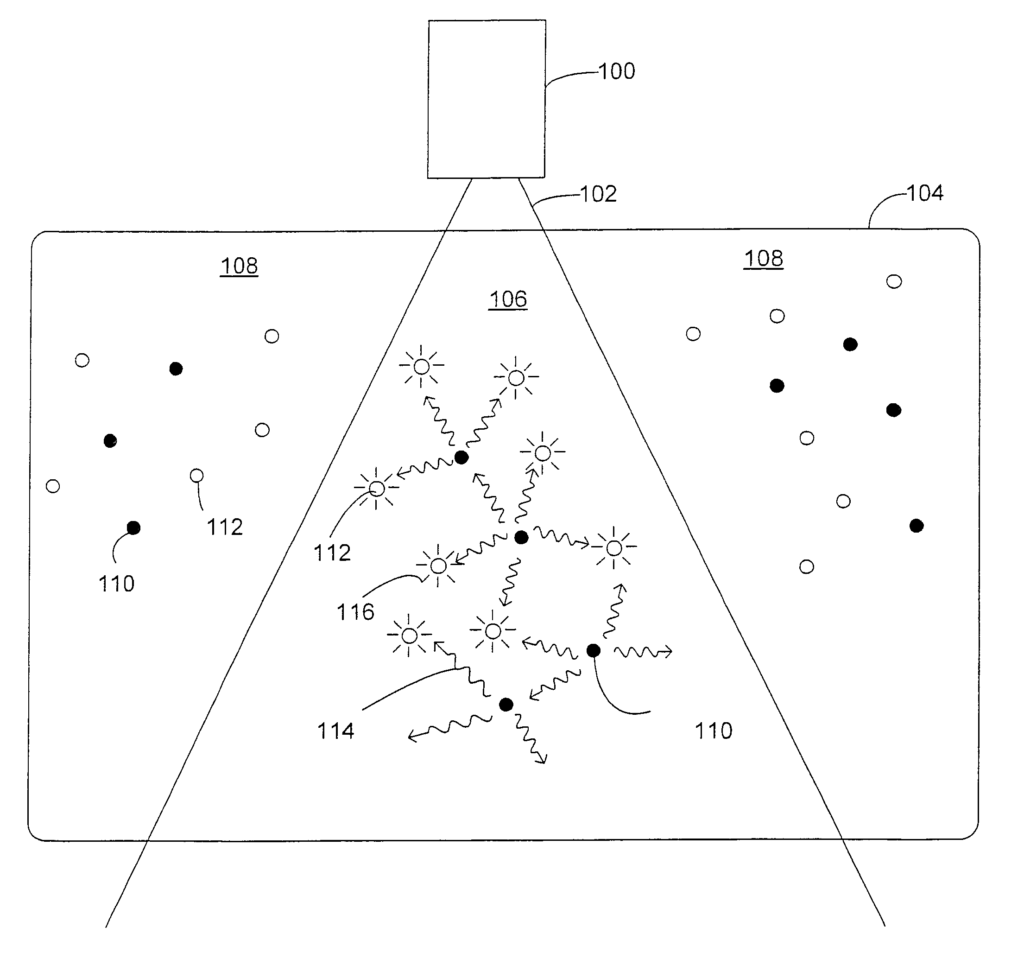
FIG. “FIG. 1 shows an example embodiment where an ionizing radio frequency emitter 100 produces an ionizing radiance 102. The ionizing rays irradiate at least a part of a region 104 containing a luminescent substance 110 and a bioactive photosensitive material 112. The region 104 could include, for instance, a patient, or a part thereof, such a the head, neck or limb of a person or an animal, or the thorax or spine, abdomen or pelvis, or a tissue, organ or gland, or any other selected area for treatment. The illustrative embodiment shown in FIG. The beam of ionizing energy divides the region into two regions: an irradiated area 106, and a non irradiated area 108. The luminescent material in the irradiated area 106 responds with optical energy to the ionizing energy 102, and the photosensitive bioactive material responds by becoming biologically active to the optical energies 114. This is shown schematically in FIG. Radial lines 116 in FIG. 1 indicate the response of the photosensitive material to the optical radiation 114. Other embodiments may provide different responses; for example the photosensitive material could respond to the light to become biologically passive, or to increase or decrease the level of biological activities, or to switch from one mode to another mode of activity. The luminescent material in the non-irradiated area 108 does not receive any ionizing radiation. Therefore, it does not produce the optical energy necessary to activate the photosensitive material.
In general, a ‘photosensitive biologically-active material’ is a term that can be used to describe any material with cellular activity. Any material with a biological activity can be included. The photosensitive biologically-active material could include, for example, materials that are biologically inactive but respond to light energy by becoming biologically active; or, materials that are biologically active that respond to light energy by becoming biologically inactive; or, materials that have a biologically low level of activity that respond to light energy in order to reach a higher level of activity.
In some embodiments, a photosensitive biologically-active material is a photographsenisitizer which responds to light by producing a reactive oxygen species such as singlet oxygen or another cytotoxic substance. Photodynamic Therapy (PDT) is a method that uses photosensitizers to destroy cancerous and diseased cells. This procedure generally involves: (1) the administration of a photosensitive drug; (2) the selective uptake or retainment of the drug in the lesion or tissue of interest; (3) the delivery of an optical light to that lesion or tissue; (4) the light absorption of the drugsensitizing agent to produce a cytotoxic substance which damages or destroys target tissue; and (5) the metabolism or excretion by the drug of the photosensitizer to reduce sunlight sensitivity. S. A. Unger’s?Photodynamic Treatment? describes photodynamic therapy, photosensitizers, and their applications in more detail. Buffalo Physician, Fall 2004, 8-19. Paras N. prasad Introduction to Biophotonics. Wiley-Interscience 2003, 433-463. Tuan Vo-Dinh, et. al. Biomedical Photonics Handbook, CRC Press 2003, 36-1-38-16. All of these are incorporated herein by reference. Porphyrins are a good example of photosensitizers. Other examples include halogenated fullerenes and dendrimers. Some applications involve administration of a metabolic precursor for a photosensitizer. An example is 5-aminolaevulinic acids (ALA), which generates endogenously the photosensitizer, photoporphyrin 9.
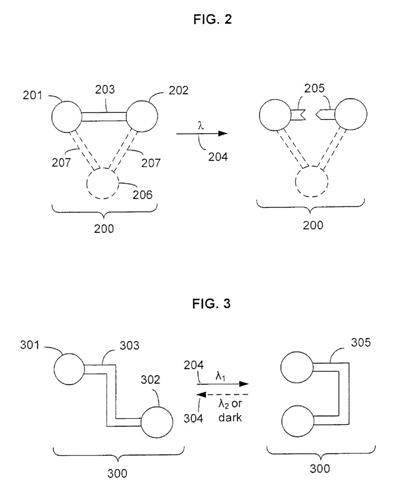
In some embodiments the biologically active photosensitive material may include a material that is photolabile. FIG. FIG. 2 shows a schematic representation of a material photolabile 200. It consists of two components 201 and 202, which are joined by a component photolabile 203. In some embodiments, the elements shown with dashed-lined are optional. As shown by the arrows 204 and 205 labeled with wavelength?, the material responds to optical energy within at least one band of wavelength to split the photolabile into two fragments. The response of the material to optical energy is described by those skilled in the art using various terms, such as?photolysis? ?photodissociation,? ?photo-release,? Photo-release,? The material can be completely cleaved if the photolabile component is the only structure connecting the first and second components. The material 200 may include a third component, 206, which is joined to the first component, 201, and the second component, 202, by non-photolabile component 207. In this case, the structure is altered in response to the optical energy within the at least one band. However, the first and second component remain in an indirect relationship. The modified or cleaved structures can have biological activities that are different from those of the unmodified and uncleaved structures.
Those skilled in the arts are aware of a variety of photosensitive biologically-active materials, including photolabile materials. Other embodiments are also obvious to those with knowledge of the art. Fay et al, ?Photosensitive caged macromolecules,? U.S. Pat. No. No. 2-nitrophenyl, 2-nitrobenzyloxycarbonyl, or ?-carboxy 2-nitrobenzyl) and responsive to optical energy to become biologically active or inactive. Grissom et. al., ‘Bioconjugates, and delivery of bioactive agent,? U.S. Pat. No. The patent 6,777,237 (herein incorporated by referencing) describes an example where a bioactive molecule is bonded to the cobalt in an organocobalt compound, and that the complex responds with light to cleave this bond, releasing the bioactive molecule. Kehayova and al., “Phototriggered Delivery of Hydrophobic Carbonic Anhydrase Inhibitors”,? Photochem. Photobiol. Sci. 1 (2002), 774-779, herein incorporated by reference, describes a carbonic anhydrase inhibitor bearing a photolabile cage compound, o-nitrodimethoxyphenylglycine (o-NDMPG) and responsive to optical light to photo-uncage (and thereby activate) the inhibitor molecule. W. Neuberger?Devices and methods for photoactivated drugs therapy? U.S. Pat. No. The patent 6,397,102 is herein incorporated as reference and describes a drug encapsulated or attached to a fullerene photolabile molecule. When the inactive drug-fullerene is exposed to selective irradiation the complex breaks down, releasing the drug in its active form. A. Momotake, et. al., “The nitrodibenzofuran: a caging group for ultra efficient photolysis in living cell,?” Nature Methods 30 (2007), 35-40 and W. H. Li, “Crafting New Cages” Nature Methods 30, (2006), 13-15 (both herein incorporated as references) describe a photolabile caging group of nitrodibenzofuran. V. Tassel et al, ?Photolytic drug delivery systems,? International Application No. PCT/US96/01333 and A. W. Lindall – “Catheter System for Controllably Releasing a Therapeutic Agent at a Remote Tissue Site” U.S. Pat. No. Nos. A 2-nitrophenyl acridine nitroaromatic arylsulfonamide or similar chromophore, responsive to optical radiation, releases the therapeutic or diagnostic agents from the substrate. Guillet et al, ?Drug delivery systems,? U.S. Pat. No. No. A photolabile peptide-blocker compound, and responsive to light in order to release the therapeutic compounds from the polymer mixture.
In some embodiments the photosensitive biologically-active material can include a material that is photoisomerizable. FIG. FIG. 3 shows a schematic representation of a material that is photoisomerizable, comprising a component 300 with a component 301 and another component 302, joined by the photoisomer in its first isomeric state 303. The material responds to optical energy within at least a certain first wavelength band as shown by the arrow 204 with wavelength?1 to convert the component into a second isomeric state 305. The figure depicts a simplified representation of isomerization. It is not meant to be restrictive. In certain embodiments, the two isomeric components of the photoisomer are trans and cis isomers. In certain embodiments, the transition from the initial isomeric to the second isomeric forms is irreversible. As indicated by the dashed line 306, in other embodiments, the transition from first isomeric to second isomeric forms is reversible. The reverse transition can occur when optical energy is present in at least one second wavelength band, as indicated by label “2”, or it may occur when there is a reduction in optical energy or an absence of optical power in at least one first wavelength band, as indicated by label “dark”. The different isomeric form of the photoisomerizable materials can have different biological activity.
Those skilled in the field are aware of a variety of photosensitive biologically-active materials, including photoisomerizable material. Other embodiments are also obvious to those with knowledge of the art. Volgraf and others, “Allosteric Control of an Ionotropic Glutamate Receptor with an Optical Switch”,? Nat. Chem. Biol. Biol. Nature Neuroscience 7, 2004: 1381-1386. Isacoff and colleagues, “Photoreactive regulators of protein function and their methods of use,” U.S. Patent Application Publication No. US2007/0128662A1, which is hereby incorporated by reference, describes photoisomerizable material responsive to light to regulate the functions of proteins. Kumita and colleagues, “Photo-control of the helix in a short polypeptide”,? PNAS 97 (2000), 3803-3808, which is hereby incorporated by reference, discusses a peptide that has been modified to include a photoisomer of azobenzene and responsive to light energy in order to increase the helix contents of the peptide.
In some embodiments, the photosensitive material biologically active includes a partner protein. The photosensitive material biologically active is responsive to light energy and can modify the interaction between the partner protein and the binding partner. Protein and binding partner could be, for instance: a receptor (e.g. An agonist, an antagonist, an inverse-agonist or a pore blocker could be the protein and binding partner. An enzyme and its ligand (e.g. an allosteric effector, inhibitor, activator, etc. Any other protein, fragment or complex of proteins, and its corresponding ligand, e.g. an element, molecule, peptide, etc.) Capable of binding to a protein, peptide, fragment or complex of proteins and then affecting their behavior. In certain embodiments, the binding partner’s probability of binding the protein is changed in response to the optical energy within the at least one band. The photosensitive biologically-active material, for example, may contain a component that is photolabile and cages or inhibits a binding partner. In response to the optical energy, this component is removed, allowing the binding partner to bind to the corresponding protein. Another example is a photoisomer. The photoisomer’s isomeric form can affect the ability of the binding partner or protein to bind. Volgraf et al., Banghart et al., and Isacoff et al., cited above, are examples of binding partners (e.g. The binding partner (e.g. a pore-blocker or receptor agonist), tethered with a photoisomer is able to move relative to the binding site due to isomerization. In other embodiments, a combination of the binding partner and the protein has a biological activity level that can be changed in response to the optical energy within the at least one band. Eisenman et. al., “Anticonvulsant effects and anesthetic effects from a fluorescent neurosteriod activated by visible lights.” Nature Neuroscience 10 (2007), 523-530, herein incorporated by reference, describes a fluorescently-tagged neurosteriod (NBD-allopregnanolone) that binds to the GABAA receptor and responds to optical light to potentiate receptor function.
In some embodiments, the photosensitive biologically active material includes a combination of a biologically active material and a photosensitive bioactivity-adjusting material, where the photosensitive bioactivity-adjusting material is responsive to optical energy to increase, decrease, or otherwise affect the biological activity of the biologically active material. For example, the photosensitive bioactivity-adjusting material may be disposed to at least partially inhibit biological activity of the biologically active material and responsive to optical energy to at least partially uninhibit biological activity of the biologically active material. Alternatively or additionally, the photosensitive bioactivity-adjusting material may be a material having a first state causing at least a first degree of inhibition of biological activity of the biologically active material and a second state causing at most a second degree of inhibition of biological activity of the biologically active material, where the first degree of inhibition is greater than the second degree of inhibition, and where the photosensitive bioactivity-adjusting material is responsive to optical energy in at least the first wavelength band to at least partially convert from an unconverted state to a converted state, the unconverted state and converted state being uniquely selected from the group consisting of the first state and the second state. In some embodiments, the conversion from the non-converted state to converted state can be irreversible. In some embodiments, the conversion of the unconverted to converted state can be reversed. The reversion (or reverse conversion) may take place in response either to an optical energy reduction in at least one second wavelength band, or to a lack or reduction in optical energy within at least one first wavelength band. The biologically-active material can include any substance with a biological or pharmaceutical effect, such as analgesics or anti-infectives; antineoplastics or other cytotoxic and chemotherapeutic drugs, cardiovascular agents or diagnostic agents; dermatological agents or EENT agents; gastrointestinal agents or gynecological or respiratory agents or agents of the urological system. A biologically active material may or may not have an intrinsic response to optical energy to change its biological activity, but the combination of a biologically active material and a photosensitive bioactivity-adjusting material can constitute a photosensitive biologically active material that is responsive to optical energy. The term “photosensitive biologically-active material” is used throughout this document. is intended to encompass materials that are a combination of a biologically active material and a photosensitive bioactivity-adjusting material, unless context dictates otherwise.

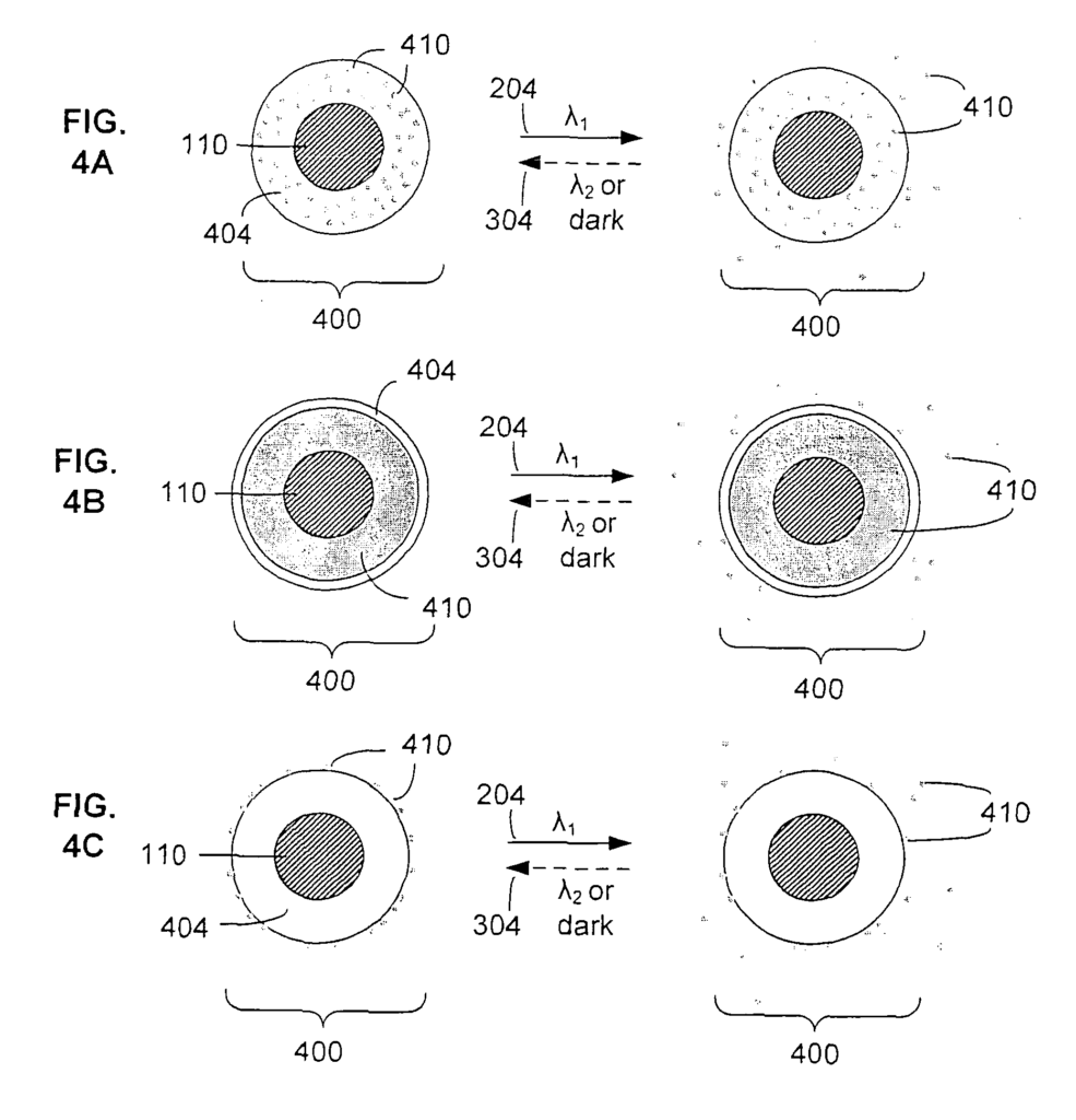
FIGS. 4A-4C depict some exemplary configurations of an ionizing-radiation-responsive composition 400 comprising a luminescent material 110, a photosensitive bioactivity-adjusting material 404, and a biologically active material 410. These configurations are only intended as examples and not to be restrictive. FIG. 4A shows a photosensitive bioactivity-adjusting material 404 disposed as a photosensitive matrix material that occupies the interstices between, or otherwise encloses, embeds, or absorbs, a plurality of portions of a biologically active material 410. FIG. 4B shows a photosensitive bioactivity-adjusting material 404 disposed as a photosensitive layer that encloses or envelops a biologically active material 410. FIB. 4C shows a photosensitive bioactivity-adjusting material 404 disposed as a substrate material having a surface that attaches, adsorbs, or otherwise couples to a biologically active material 410. The FIGS. The core-shell structures of FIGS. 4A-4C are shown with a core 110 of luminescent materials, but the disposition of this material is only intended as an example and not to be taken as a limitation. In other embodiments of the ionizing-radiation-responsive composition 400, the luminescent material is unattached to either the biologically active material or the photosensitive bioactivity-adjusting material, at least partially attached to one or the other, or variously disposed in configurations that combine all three materials. Some configurations of an ionizing that combine a luminescent material and a photosensitive biologically active material (where the latter may itself comprise a biologically active material and a photosensitive bioactivity-adjusting material) are described elsewhere. In each of FIGS. 4A-4C, the photosensitive bioactivity-adjusting material is responsive to optical energy in at least a first wavelength band, as depicted by the arrow 204 labeled with a wavelength ?1, to at least partially allow release of the biologically active material 410. In some embodiments, the response to the optical energy is irreversible. In other embodiments, the response is reversible as shown by the dashed line 304 illustrating a reversion. The reversion can occur as a result of optical energy in a second band at least (as shown by the label “2”) or in response to an absence or reduction of optical energy within the first band at least (as shown by the label “dark ?).”).
In some embodiments, the photosensitive bioactivity-adjusting material may include a substrate material having a surface that attaches, adsorbs, or otherwise couples to a biologically active material, and responsive to optical energy to release the biologically active material from the surface (optionally, embodiments include a linking agent, e.g. A bifunctional photolytic linking agent, which connects the substrate and biologically-active material, responds to light energy by separating the two materials, for example, Photolysis is one example of a photolytic linker. Examples of materials that may be used in embodiments include those described in Van Tassel, et. al. and Lindall (both previously cited and incorporated herein by reference). Various substrate materials include natural polymers, synthetic polymers, silica, glass, quartz, metal, and any other materials capable of directly or indirectly binding to the biologically active material (in some embodiments the luminescent material, or another constituent of the ionizing-radiation-responsive composition, may serve as the substrate material). “Various linking agents can include 2-nitrophenyl group, acridines or nitroaromatics or arylsulfonamides or other photolytic agents that are capable of binding directly or indirectly to the substrate and biologically active material.
In some embodiments, the photosensitive bioactivity-adjusting material includes a material that responds to optical energy to change a diffusion characteristic of the material, which may affect a rate of diffusion of the biologically active material through the photosensitive bioactivity-adjusting material. Examples of embodiments include materials like those described in Fink and colleagues, “Photoactivated Drug Therapy.” U.S. Patent Application Publication No. U.S. Patent Application Publication No.
In some embodiments, the photosensitive bioactivity-adjusting material may include a material that responds to optical energy to undergo a shape change (e.g. The shape change can be an expansion, contraction or bending; the shape-change may correspond to a different diffusion characteristic or it may affect another way of releasing the biologically active materials (e.g. A shrinkage can create a force that releases the biologically-active material or a bend may release it through a gate. As an example, embodiments can use materials like those described in Rosenthal et. al., “Triggered Release Hydrogel Drug Delivery System”. U.S. Pat. No. This reference describes catheters with a polymer gel or polymer disposed to immobilize and incorporate a drug and that responds to optical light by contracting or swelling to release the drug. Embodiments may use a light-sensitive copolymer or copolymer gel, where a first component of the light-sensitive copolymer or copolymer gel is polyacrylamide, poly(N-isopropylacrylamide), hydroxyethyl methacrylate, dihydroxypropyl methacrylate, a copolymer or mixture thereof, or the like, and a second component of the light-sensitive copolymer or copolymer gel is a light-sensitive compound that induces swelling (as with malachite green derivatives, leucocyanides, leucohydroxides, or similar compounds, e.g. As described in “Photoinduced phase change of gels”, Macromolecules 23, 1990, 1517-1519 (herein incorporated as a reference) and Guillet et. al., supra. Phase transitions in polymer gels are induced by visible lights, as described in “Phase Transition in Polymer Gels Induced By Visible Light” Nature 346 (1990), pp. 345-347 (here incorporated by renvoi) the copolymer or gel that is sensitive to light in response to optical energy.
In some embodiments, the photosensitive bioactivity-adjusting material may include a material that responds to optical energy to at least partially photodegrade, photodissociate, or pholodisintegrate (such terms may be used interchangeably); the photodegradation, photodissociation, or photodisintegration may correspond to a change of a diffusion characteristic, or affect some other means for release of the biologically active material (e.g. a mechanical disintegration of the photosensitive bioactivity-adjusting material may cause an exposure or dispersal of the biologically active material), or both. As an example, embodiments can use photochemically-degradable polymers, such as those described by Guillet et al., supra (e.g. “Copolymers of unsaturated monomers and ethylenically unsaturated monomers.
In some embodiments, the photosensitive bioactivity-adjusting material may include a material that responds to optical energy to change its hydrophobicity, hydrophilicity, or amphiphilicity; this change may correspond to a change of a diffusion characteristic, or affect some other means for release of the biologically active material (e.g. The change could cause a separation of hydrophilic components and hydrophobic materials that are immiscible, or both. As an example, embodiments can use polymers which convert photochemically between a hydrophobic and a hydrophilic form. These are described by Guillet et al., supra (e.g. Polymers that include a tbutylketone group adjacent to the backbone of the polymer.
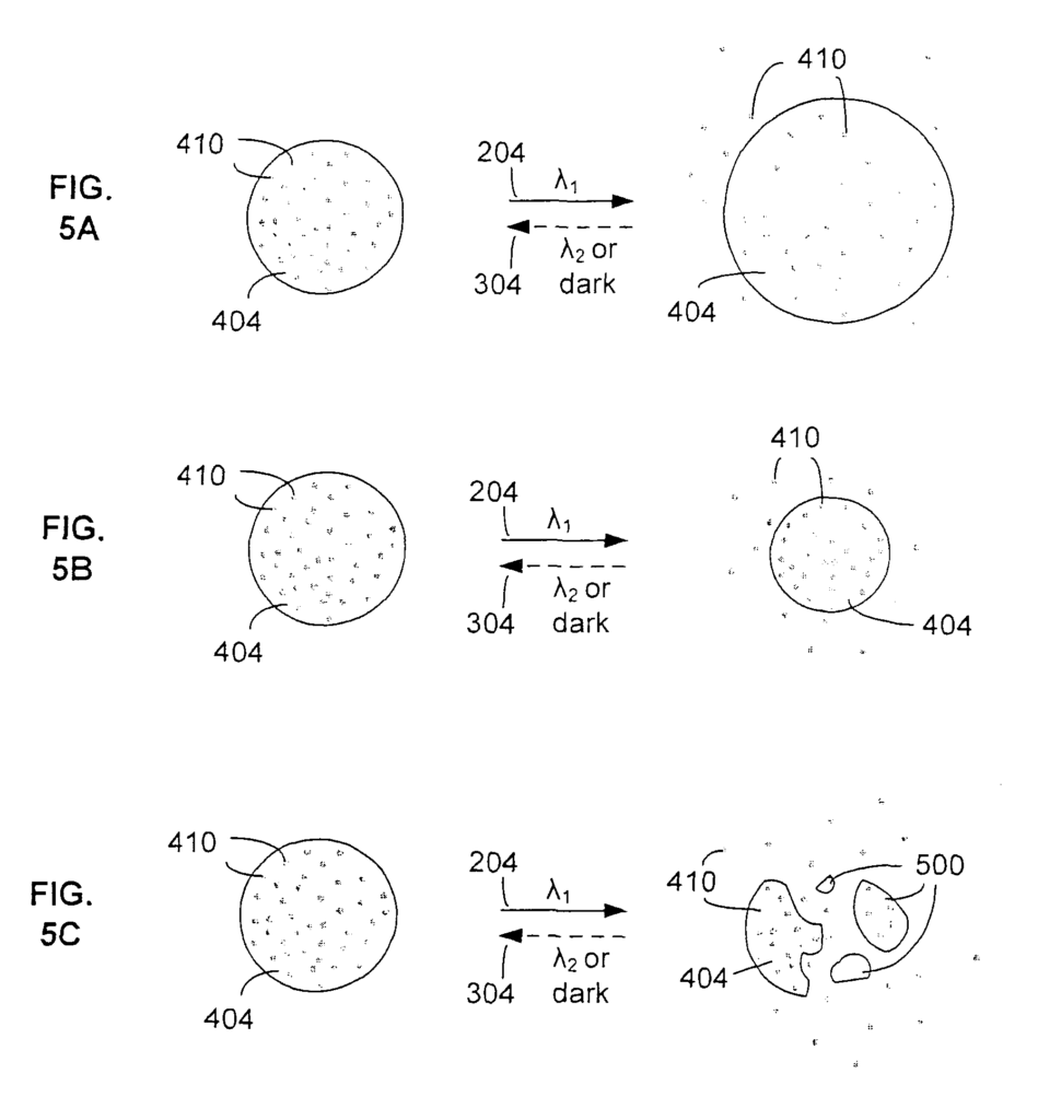
Referring now to FIGS. 5A-5C, some illustrative examples of the preceding embodiments are shown, including a photosensitive bioactivity-adjusting material 404 and a biologically active material 410. For purposes of clarity, a luminescent material is not depicted in these examples, but this omission is not intended to be limiting, and embodiments provide a luminescent material that is enclosed, attached, or otherwise disposed in a vicinity of the photosensitive bioactivity-adjusting material and/or the biologically active material. FIG. 5A depicts an example of a photosensitive bioactivity-adjusting material 404 disposed as a photosensitive matrix material enclosing a biologically active material 410, and responsive to optical energy in at least a first wavelength band (as depicted by the arrow 204 labeled with a wavelength ?1) to expand, the expansion causing a release (e.g. The biologically active material is released (e.g. FIG. 5B depicts an example of a photosensitive bioactivity-adjusting material 404 disposed as a photosensitive matrix material enclosing a biologically active material 410, and responsive to optical energy in at least a first wavelength band (as depicted by the arrow 204 labeled with a wavelength ?1) to contract, the contraction causing a release (e.g. The biologically active material is released (e.g. by pressure expulsion). In relation to FIG. The photosensitive matrix may initially be disposed so that it allows at least partial release (e.g. The photosensitive matrix material may be initially disposed to at least partially allow release (e.g. By reducing diffusion of the biologically-active material. FIG. 5C depicts an example of a photosensitive bioactivity-adjusting material 404 disposed as a photosensitive matrix material enclosing a biologically active material 410, and responsive to optical energy in at least a first wavelength band (as depicted by the arrow 204 labeled with a wavelength ?1) to at least partially photodegrade, photodissociate, or photodisintegrate, thereby releasing the biologically active material 410 (and optionally releasing fragments 500 of the photosensitive bioactivity-adjusting material). In some embodiments, the process shown in FIGS. The process depicted in FIGS. 5A-5C may be irreversible. In other embodiments, the process can be reversed as shown by the dashed line 304 depicting the reverse process. The reverse process can occur when optical energy is detected in at least a 2nd wavelength band, as indicated by label?2, or it may happen in response to an absence or reduction of optical energy within at least 1 wavelength band.
In some embodiments, the photosensitive bioactivity-adjusting material may include a photosensitive layer (or a plurality thereof) disposed to at least partially enclose or envelop at least a portion of the biologically active material, and responsive to optical energy to at least partially allow release of the biologically active material. The term “layer” is used. The term?layer’ is meant to include a wide range of structures, including membranes and films, coatings, covers, patches, shells etc. The term “layer” is used. The term?layer’ also includes micelles, liposomes and lipid membranes as well as other monolayers or bilayers. Assembled from phospholipids or amphiphilic block polymers. In certain embodiments, the photosensitive layer can include one or more of the materials described above, e.g. A material that changes its diffusion characteristics in response to optical energy, or a substance that undergoes a change of shape (e.g. A material that is responsive to optical energy and undergoes an expansion, contraction or bending, a substance that is responsive to optical energies that at least partially photodegrades, photodissociates, or photosintegrates (thereby disrupting, perforating or otherwise disrupting the material), or a substance that is responsive to optical energies that change its hydrophobicity or hydrophilicity. A photosensitive layer may be used to embed one or more light sensitive channel proteins, such as those described by Kocer et. al., “A light-actuated Nanovalve derived From a Channel Protein.” Science 309, 2005, 755-758 and Kocer, et. al., “Modified MscL Protein Channel”, U.S. Patent Application Publication No. US2006/0258587 and US2006/0258587 are both incorporated herein by reference. These references describe a modified membrane channel protein that is responsive to an optical energy in order to irreversibly (or reversibly) open or close a pore within the membrane. Other embodiments can use materials like those described in P. Ball’s?Light Pumps Drugs from Nanoparticles? Nanozone News, Jun. 9, 2005, herein incorporated by reference; e.g., a liposomal membrane (or similar monolayer/bilayer/etc.) Photoisomerizable Amphiphiles or phospholipids are at least partially present in the membrane. Membrane proteins are integral to the membrane if it is at least partially composed of photoisomerizable phospholipids (or similar photoisomizable amphiphiles) or incorporates cholesterol that can be photoisomerizable. Open pores, rupture or perforation or other release of biologically active material.
Referring now to FIGS. 6A-6C, some illustrative examples of the preceding embodiments are shown, including a biologically active material 410 and a photosensitive bioactivity-adjusting material 404 disposed as a photosensitive layer that encloses the biologically active material. For purposes of clarity, a luminescent material is not depicted in these examples, but this omission is not intended to be limiting, and embodiments provide a luminescent material that is enclosed, attached, or otherwise disposed in a vicinity of the photosensitive bioactivity-adjusting material and/or the biologically active material. FIG. FIG. 6A shows an example of how a photosensitive film that responds to optical energy can rupture or perforate in at least one wavelength band (as shown by the arrows 204, labeled as wavelength?1), releasing the biologically active substance through the ruptured areas or perforated regions 600. FIG. FIG. 6B shows an example of a layer embedded with one or more porelike structures (e.g. The channel proteins are shown in a closed configuration 602. They respond to the optical energy within a certain wavelength range (as indicated by the arrow 204 with the wavelength?1) and convert into an open configuration 604. FIG. FIG. 6C shows an example of a layer with embedded photoisomerizable molecule (e.g. Photoisomerizable cholesterols or phospholipids are shown in a photosensitive layer 606 in a photosensitive first isomeric state. The molecules can be converted to a second form 608 by using optical energy within a certain wavelength range (as indicated by the arrows 204 with wavelength?1), thereby altering the diffusion or porosity of the photosensitive material. In some embodiments, the process shown in FIGS. The process depicted in FIGS. 6A-6C may be irreversible. In other embodiments, the process can be reversed as shown by the dashed line 304 that shows a reversed process. The reverse process can occur when optical energy is detected in at least a 2nd wavelength band, as indicated by label?2, or it may happen in response to an absence or reduction of optical energy within at least 1 wavelength band.
Treating tissue or a lesion with a biologically active photosensitive material” typically involves local irradiation of the tissue or area with optical light. The term optical light or energy is used to describe electromagnetic radiation that falls within the visible portion (e.g. having wavelengths in the range of 380 nm to 750 nm or frequencies in the range of 400 to 800 THz) as well as neighboring regions of the electromagnetic spectrum (including but not limited to far-infrared, infrared, near-infrared, near-ultraviolet, ultraviolet, and extreme-ultraviolet). The term ‘optical light’ is used. The terms ‘optical light? also encompass quantized electromagnetic radiation (i.e. Photons and other forms of non-radiative electromagnetic energy are also included (e.g. Standing waves, evanescent field, Forster resonance (FRET) energy transfer, etc.). The red and near infrared spectrum of light (the most penetrational) can penetrate up to 6mm depending on wavelength and tissue. Existing therapies are limited by the difficulty of delivering light into non-superficial regions. This is often achieved through interstitial, intravascular, or intracavitary placement of optical fibres with diffuser tips coupled to a light source. “Some embodiments provide an alternative mode of optical delivery whereby the optical light is emitted locally by a luminescent substance in response to ionizing light, which can be highly penetrational and precisely delivered to the region of interest.

Radiation that can ionize a molecule or an atom is called “ionizing radiation”. Radiation can be referred as ionizing whether it causes ionization or not in any embodiment or application of the aspects described. Ionizing radiation, for example, may have an energy that is sufficient to ionize one type of atom, but not enough to ionize another. In some embodiments, the ionizing energy may only interact with the second type of atom or molecular. “The ionizing radiation may be electromagnetic radiation, such as soft or hard x rays or gamma rays or charged particle radiation, in the form electrons or protons (e.g. carbon and neon).